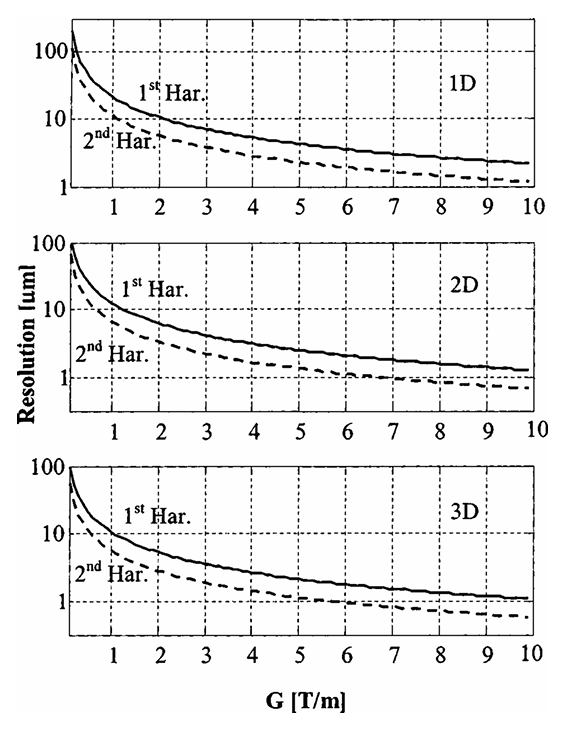
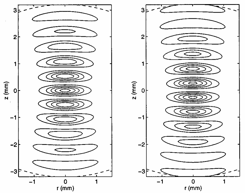
ABSTRACT: An electron spin resonance (ESR) imaging system, capable of acquiring three-dimensional (3D) images with a resolution of ˜10 × 10 × 30 μm in a few minutes of acquisition, is presented. This ESR microscope employs a commercial continuous wave ESR spectrometer, working at 9.1 GHz, in conjunction with a miniature imaging probe (resonator + gradient coils), gradient current drivers, and control software. The system can acquire the image of a small (˜ 1.5 × 1.5 × 0.25 mm) sample either by the modulated field gradient method, the projection reconstruction method, or by a combination of the two. A short discussion regarding the resolution of the modulated field gradient method in two-dimensional (2D) and 3D imaging is given. Detailed descriptions of the various system components are provided, along with several examples of 2D and 3D images that demonstrate the capabilities of the system.
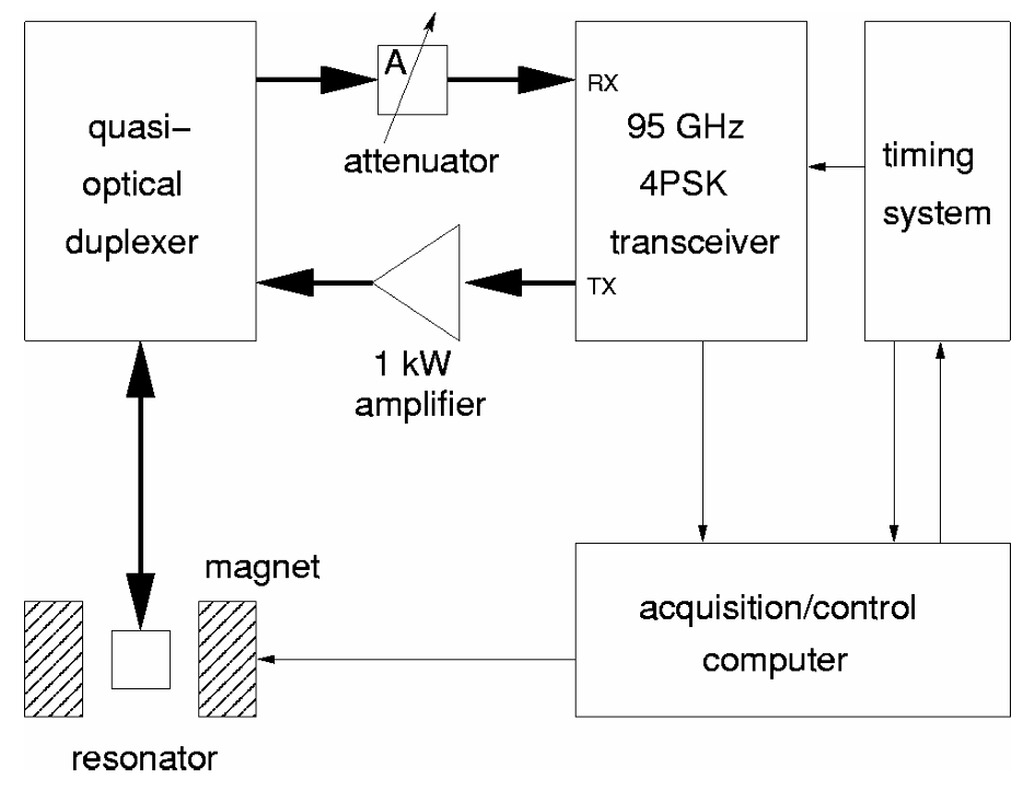
ABSTRACT: High-field/high-frequency electron spin resonance (ESR) offers improved sensitivity and resolution compared to ESR at conventional fields and frequencies. However, most high-field/high-frequency ESR spectrometers suffer from limited mm-wave power, thereby requiring long mm-wave pulses. This precludes their use when relaxation times are short, e.g., in fluid samples. Low mm-wave power is also a major factor limiting the achievable spectral coverage and thereby the multiplex advantage of Fourier transform ESR (FTESR) experiments. High-power pulses are needed to perform two-dimensional (2D) FTESR experiments, which can unravel the dynamics of a spin system in great detail, making it an excellent tool for studying spin and molecular dynamics. We report on the design and implementation of a high-power, high-bandwidth, pulsed ESR spectrometer operating at 95 GHz. One of the principal design goals was the ability to investigate dynamic processes in aqueous samples at physiological temperatures with the intent to study biological systems. In initial experiments on aqueous samples at room temperature, we achieved 200 MHz spectral coverage at a sensitivity of 1.1×10¹⁰√(s) spins and a dead time of less than 50 ns. 2D-electron-electron double resonance experiments on aqueous samples are discussed to demonstrate the practical application of such a spectrometer.
ABSTRACT: We report on the performance of a Fabry–Perot resonator for far-infrared electron spin resonance (FIR-ESR) at 250 GHz designed to accommodate a thin, disk-shaped sample that must rest with its flat surface perpendicular to the incident FIR beam. This geometry minimizes dielectric losses, making it possible to obtain FIR-ESR spectra of aqueous or lossy samples with a macroscopic ordering, at canonical values of the director tilt of 0° and 90°. The resonator also utilizes an adjustable interferometer to achieve variable coupling in the FIR regime.
ABSTRACT: A quasioptical approach is utilized to design a resonator and sample holder for aqueous samples for high-frequency (250 GHz) electron spin resonance (ESR) spectroscopy. A disk shaped sample geometry was chosen to match the field contours of the fundamental mode in a Fabry–Perot resonator. A transmission-line analysis is used to determine the optimum sample geometry while taking into account diffractive effects of the sample and the sample holder on the far infrared (FIR) radiation field. FIR-ESR spectra of several aqueous solutions of nitroxide spin labels, including membrane vesicles and a labeled protein, are shown to demonstrate the success of the technique.
ABSTRACT: We describe a far-infrared electron-paramagnetic-resonance (EPR) spectrometer for broadband (100–300 GHz) use. The spectrometer is operated in the reflection mode and uses broadband quasioptical methods to separate the transmitted from the reflected radiation. We describe and illustrate its operation at 170 GHz (1.8 mm) and compare its performance to that of our transmission mode spectrometer operating at 250 GHz. We also discuss the advantages of the reflection bridge for performing EPR experiments over a broad range of frequencies, and we consider methods of improving the performance of the bridge. This includes a novel design for variable coupling of the reflection-mode Fabry–Pérot resonator.
ABSTRACT: An ESR spectrometer operating at 250-GHz frequency (1.22-mm wavelength) and 9-T magnetic field is described. The utilization of far-infrared (FIR) technology greatly simplifies its design and performance. Good frequency and field stability are achieved by unique designs which also conveniently permit the magnetic field to be swept. The potential utility of FIR–ESR is illustrated with examples of spectra from polycrystalline and liquid samples. In the latter case the increased spectral sensitivity to motional dynamics is stressed. Several ways in which the FIR–ESR spectrometer may be improved are also discussed.
ABSTRACT: A method is described whereby Carr–Purcell (CP) multipulse sequences may be generated at a rate rapid enough for electron-spin echoes ESE and in a manner consistent with constraints of typical ESE spectrometers. Both Carr–Purcell–Meiboom–Gill and alternating phase-CP sequences may be produced. It is shown how such sequences may be utilized (1) to increase the rate of data collection, which is especially important in laser photo-induced experiments, and (2) to study slow motions. They are respectively illustrated with results on transient spin-polarized photoelectrons and electrons trapped in irradiated quartz.
.svg)